The typical nude skin cancer checks with long photo sessions at your dermatologist’s office to track any suspicious skin marks just got a lot more accurate, while reducing the chance of unnecessary biopsies.
UConn Health is the only institution to date in Connecticut to offer the latest advanced smart technology that hunts for skin cancer and keeps an eye on changing moles.
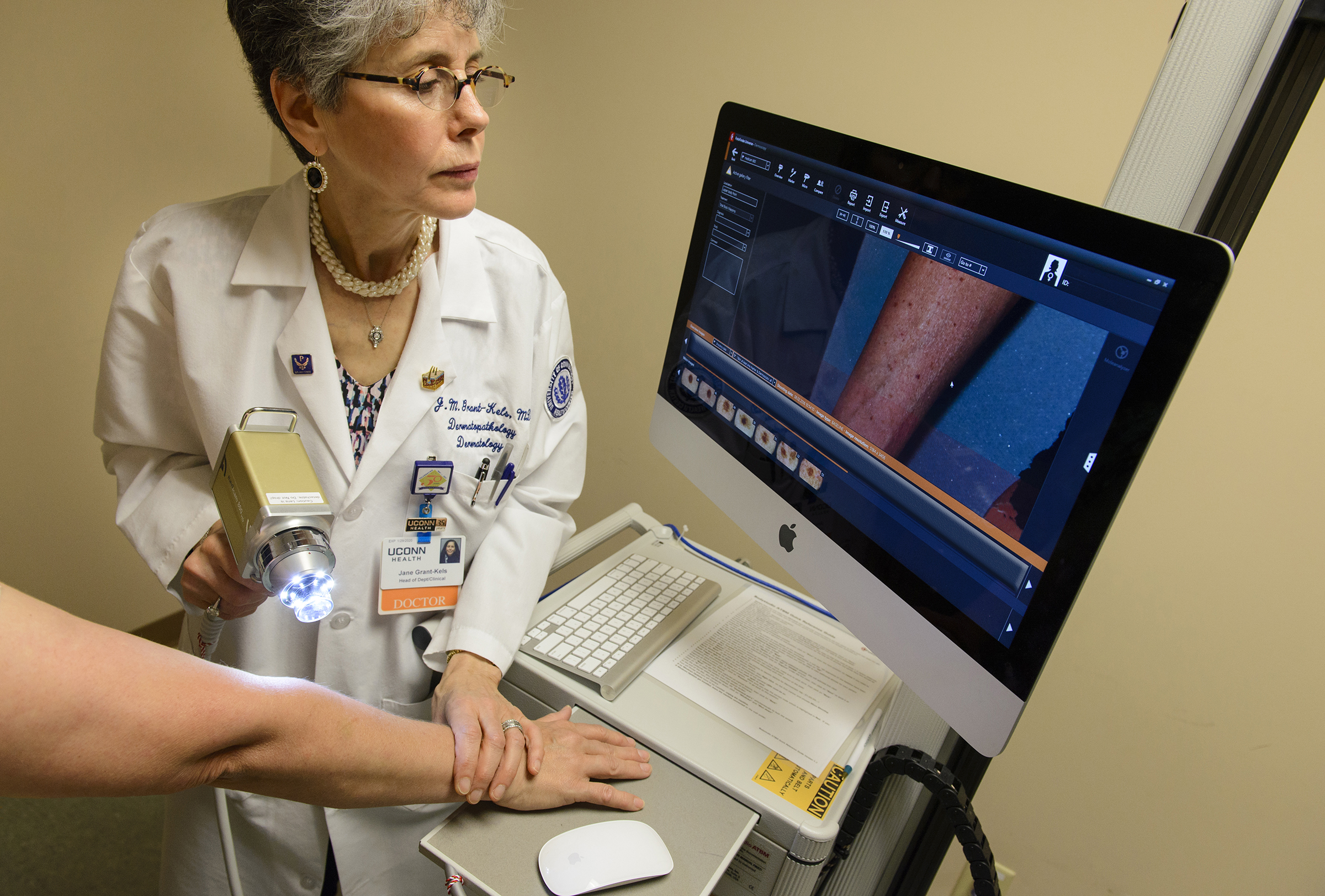
An integrated body-scanning camera and smart software technology “helps us find skin cancer in a flash,” says Dr. Jane Grant-Kels, professor and vice chair of UConn Health’s Department of Dermatology and director of the UConn Cutaneous Oncology Center and Melanoma Program.
The technology, called FotoFinder Bodystudio Automated Total Body Mapping, allows dermatology staff to take 20 or more photos of a patient’s entire body, including the palms and the soles of the feet, in about 10 minutes. It also allows easy comparison of photographs visit after visit, and alerts the dermatologist to changes or new growths.
“This technology is going to help us save more lives from skin cancer and melanoma,” says Grant-Kels. “It allows for early detection and a more exact science of monitoring patients’ skin changes.”
If concerning growths are detected, another recently arrived technology called In Vivo Reflectance Confocal Microscopy uses a non-invasive optical imaging technique that provides a high-resolution cellular image of the skin. This new technology is safe and painless, and in many cases can be used in lieu of a painful skin biopsy.
“FotoFinder coupled with Confocal will help us go a long way to reducing the number of biopsies performed, including unnecessary biopsies of non-cancerous skin growths,” Grant-Kels says.
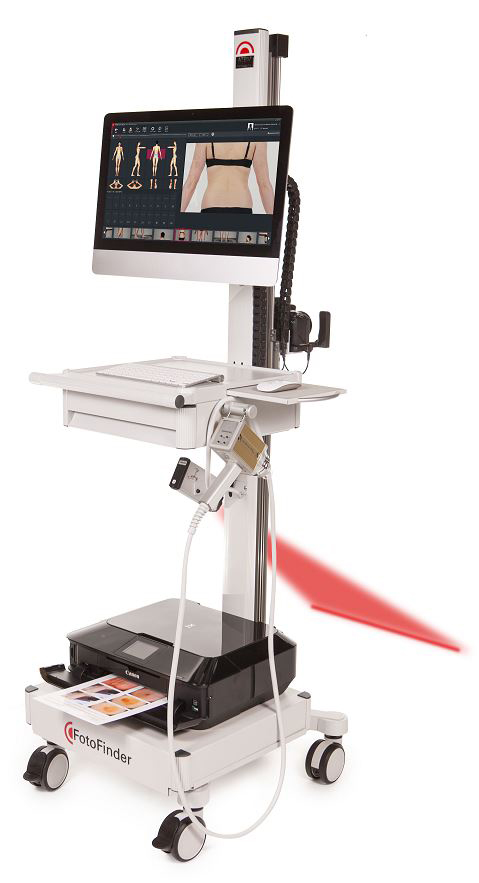
For baseline and follow-up photo sessions using the FotoFinder technology, a patient will be guided by a red laser light and a specially designed floor mat to ensure proper positioning. The smart body-scanning camera automatically moves into various positions to take photos of the entire body, and the software module rapidly stitches the photos together for the dermatologist to review.
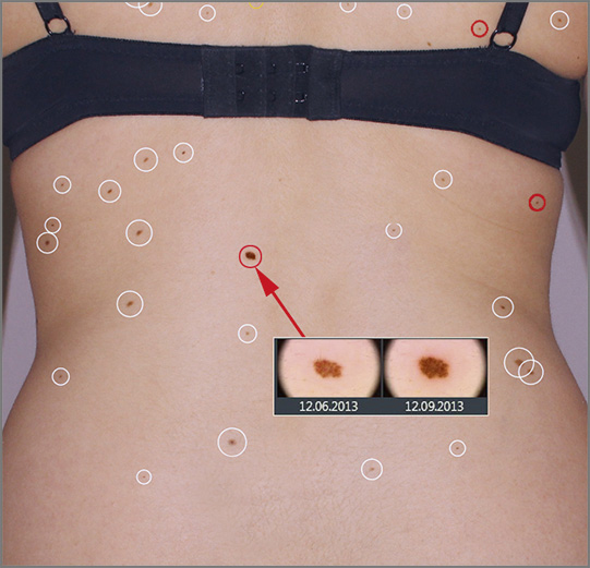
After the patient’s follow-up photo session, within seconds the system uses its computer technology to precisely place the most recent skin images atop the baseline photos. The software seamlessly aligns and analyzes the new and old photos, and then circles all the detected new and visibly changed skin lesions and moles.
White circles around lesions or moles signal to the dermatologist no change; yellow circles signal caution to the doctor, as the lesion or mole has changed since the last visit; and red circles raise alarm for the doctor, as a new lesion or mole growth has been identified. This allows the dermatologist to investigate the most alarming skin lesions first.
The technology also allows dermatologists to compare lesion or mole photos side by side and to quickly zoom from 20x- up to 70x-magnification to examine suspicious areas in high-resolution and determine which spots to examine more closely with the traditional handheld dermoscopy tool. The system also includes high-tech, handheld electronic dermoscopy with a built-in medicam for even closer examination and additional photo captures. Plus, the machine is mobile and can be moved easily among exam rooms.
Grant-Kels recommends that patients with many moles or a family history of skin cancer or melanoma see a dermatologist every six months.
“I am glad to report that UConn Health now has every new FDA-approved technology to help patients manage skin health, placing us on the cutting-edge of skin cancer prevention, detection, and treatment,” she says.
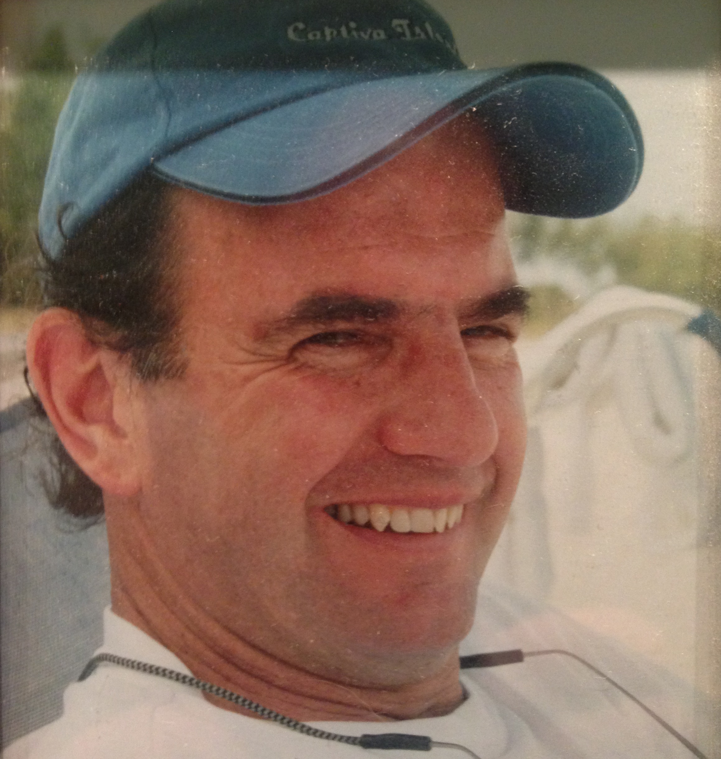
“I am at high-risk for skin cancer, since I have like 400,000 moles,” jokes Pierre Simler, 69, of Litchfield, Conn. Also, as a military veteran he spent a lot of time outside in the sun during his service.
Simler is a three-time skin cancer survivor and has had more than a dozen moles biopsied and removed.
But now Simler’s annual skin checks and biopsy worries are way less burdensome, thanks to FotoFinder technology at UConn Health.
The new integrated body-scanning camera and smart software technology is helping Simler’s UConn Health dermatologist, Dr. Marti Rothe, find any suspicious lesions or changing moles in about 10 minutes, and easily track his skin changes over time by comparing photographs visit after visit.
“It is really high-tech and unbelievable,” says Simler. “The machine takes my skin photos, and during the next visit my doctor can quickly tell exactly what mole has changed, plus the computer tells her which one is most critical to look at first.”
If a suspicious lesion or mole is identified, Confocal technology is then used to take a deeper, non-invasive look into the skin, leveraging the power of painless laser light. This technology can allow dermatologists to avoid a biopsy or mole removal if the closer look of the skin’s cells using Confocal doesn’t show warning signs of cancer.
Adds Simler, “To me a doctor’s eye is great. But a doctor’s eye plus this new technology is even better.”
Dermatologists at UConn Health believe that, with this Fotofinder and Confocal technology advancements, more people will feel more comfortable coming in for their skin checks, knowing their skin lesion or mole is not just immediately going to be biopsied or cut out.
Simler is now adamant about wearing 70 SPF sunscreen, t-shirts, and a hat when outdoors, and gets a skin check every six months. “A tan is not cool,” he stresses.
He recommends that everyone does the same.
“You have no idea if you truly have a hereditary risk for skin cancer,” he says. “Even if you take precautions and stay out of the sun, you can still get skin cancer. A skin check needs to become part of your annual medical routine. It could save your life.”
If you have a lot of moles or a family history of skin cancer or melanoma, the dermatologists at UConn Health recommend you should be seeing your dermatologist every six months.
To learn more about dermatology services at UConn Health visit: http://health.uconn.edu/dermatology/