In the not so distant past, medical and dental students would learn minimal amounts of radiology and medical imaging along with their gross anatomy experience with cadavers. But now UConn Health’s Virtual Anatomy Lab, better known as the “VAL,” greatly expands that experience. It allows first- and second-year medical and dental students to solidify their learning of anatomy along with introducing them to important new imaging technology they’ll be using throughout their careers. Step into the lab to observe first-hand how the students – and ultimately their patients – benefit from what the VAL has to offer.
In the ‘VAL’ – Learning the Virtual Way
UConn Health’s Virtual Anatomy Lab, the “VAL,” allows medical and dental students to solidify their learning of anatomy along with introducing them to new imaging technology. Step into the lab to observe first-hand how the students – and ultimately their patients - benefit from what the VAL has to offer.
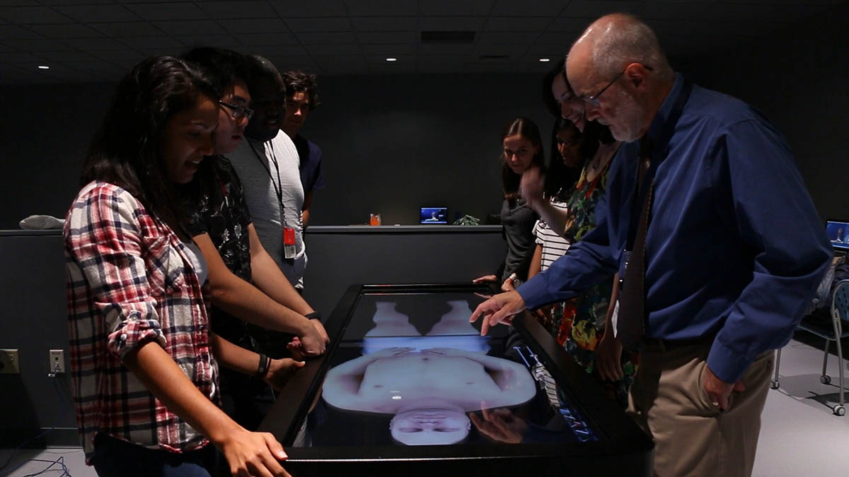
John Harrison, the director of the Human and Virtual Anatomy Lab at UConn Health, instructs medical and dental students on the use of the Anatomage table in the Virtual Anatomy Lab (VAL). (Frank Barton/UConn Health)