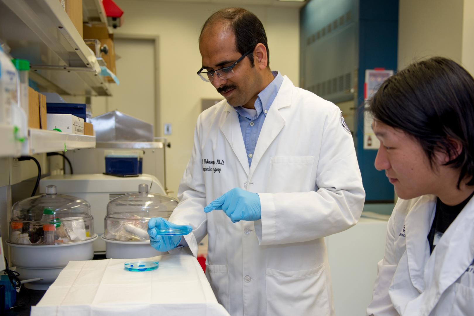
A team of UConn Health researchers has designed a novel, hybrid hydrogel system to help address some of the challenges in repairing bone in the event of injury. The UConn Health team, led by associate professor of orthopedic surgery Syam Nukavarapu, described their findings in a recent issue of Journal of Biomedical Materials Research-Part B, where the work is featured on the journal cover.
There are over 200 bones in an adult human skeleton, ranging in size from a couple of millimeters in length to well over a foot. How these bones form and how they are repaired if injured varies, and has posed a challenge for many researchers in the field of regenerative medicine.
Two processes involved with human skeletal development help all the bones in our body form and grow. These processes are called intramembranous and endochondral ossification, IO and EO respectively. While they are both critical, IO is the process responsible for the formation of flat bones, and EO is the process that forms long bones like femurs and humeri.
For both processes, generic mesenchymal stem cells (MSCs) are needed to trigger the growth of new bone. Despite this similarity, IO is significantly easier to recreate in the lab since MSCs can directly differentiate, or become specialized, into bone-forming cells without taking any additional steps.
However, this relative simplicity comes with limitations. To circumvent the issues associated with IO, the UConn Health team set out to develop an engineered extracellular matrix that uses hydrogels to guide and support the formation of bone through EO.
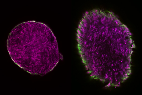
“Thus far, very few studies have been focused on matrix designs for endochondral ossification to regenerate and repair long bone,” says Nukavarapu, who holds joint appointments in the departments of biomedical engineering and materials science and engineering. “By developing a hybrid hydrogel combination, we were able to form an engineered extracellular matrix that could support cartilage-template formation.”
Nukavarapu notes that vascularization is the key in segmental bone defect repair and regeneration. The main problem with IO-formed bone is caused by a lack of blood vessels, also called vascularization. This means that IO isn’t capable of regenerating enough bone tissue to be applied to large bone defects that result from trauma or degenerative diseases like osteoporosis. Although many researchers have tried various strategies, successfully vascularizing bone regenerated with IO remains a significant challenge.
On the other hand, vascularization is a natural outcome of EO due to the development of a cartilage template, chondrocyte hypertrophy, and eventual bone tissue formation.
While IO’s simplicity caused limitations, EO’s benefits result in an intricate balancing act. EO requires precise spatial and temporal coordination of different elements, like cells, growth factors, and an extracellular matrix, or scaffold, onto which the MSCs attach, proliferate, and differentiate.

To achieve this delicate balance in the lab, Nukavarapu and his colleagues combined two materials known to encourage tissue regeneration – fibrin and hyaluronan – to create an effective extracellular matrix for long bone formation. Fibrin gel mimics human bone mesenchymal stem cells and facilitates their condensation, which is required for MSC differentiation into chondrogenic cells. Hyaluronan, a naturally occurring biopolymer, mimics the later stages of the process by which differentiated chondrogenic cells grow and proliferate, also known as hypertrophic-chondrogenic differentiation.
The researchers anticipate that cartilage templates with hypertrophic chondrocytes will release bone and vessel forming factors and will also initiate vascularized bone formation. Nukavarapu says that the “use of cartilage-template matrices would lead to the development of novel bone repair strategies that do not involve harmful growth factors.”
While still in the early research phase, these developments hold promise for future innovations.
“Dr. Nukavarapu’s work speaks not only to the preeminence of UConn’s faculty, but also to the potential real-world applications of their research,” says Radenka Maric, vice president for research at UConn and UConn Health. “UConn labs are buzzing with these types of innovations that contribute to scientific breakthroughs in healthcare, engineering, materials science, and many other fields.”
The researchers next plan to integrate the hybrid extracellular matrix with a load-bearing scaffold to develop cartilage templates suitable for long-bone defect repair. According to Nukavarapu, the UConn research team is hopeful that this is the first step towards forming a hypertrophic cartilage template with all the right ingredients to initiate bone tissue formation, vascularization, remodeling, and ultimately the establishment of functional bone marrow to repair long bone defects through EO.
The work was supported by grants from the AO Foundation (S-13-122N), NSF Emerging Frontiers in Research and Innovation (EFRI) (1332329), and NSF Emerging Frontiers and Multidisciplinary Activities (EFMA) (1640008).
Research in Syam Nukavarapu’s lab focuses on biomaterials and tissue engineering, with emphasis on bone, cartilage, and bone-cartilage interface tissue engineering. Other UConn authors include graduate students Paiyz E. Mikael and Hyun S. Kim.