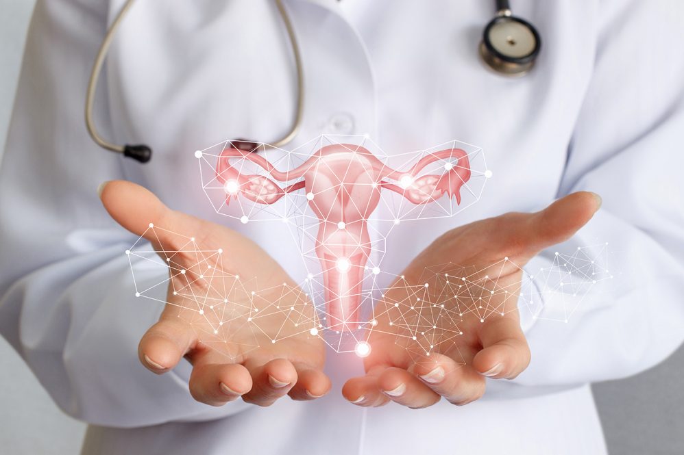
The tissue that lines the uterus, known as the endometrium, serves as the location of embryo implantation and the source of the arteries that lead into the placenta to support a fetus during pregnancy. But in humans, when there is no fertilized egg, the endometrium is shed through menstruation. The endometrium is thus unusual in that it is regularly lost, then proliferates again, throughout a woman’s reproductive age. In about 10 percent of women, however, endometrium-like tissues (known as lesions) also grow outside of the uterus, leading to endometriosis.
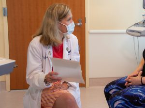
Endometriosis is characterized by pain and can cause infertility, but its molecular mechanisms and drivers remain unknown. The Jackson Laboratory’s (JAX) Elise Courtois, Ph.D., is leading research efforts to reveal what causes endometriosis and to focus more clinical attention on it in partnership with UConn Health’s gynecological surgeon Danielle Luciano, M.D.
Definitive diagnosis and clinical response still present significant challenges, with a common treatment being hormonal therapy with surgery. Unfortunately, surgery must be repeated if lesions recur, and they often do. To improve the situation, a better understanding of how and why the lesions grow, their cellular makeup, their microenvironments, and other aspects of their biology is essential.
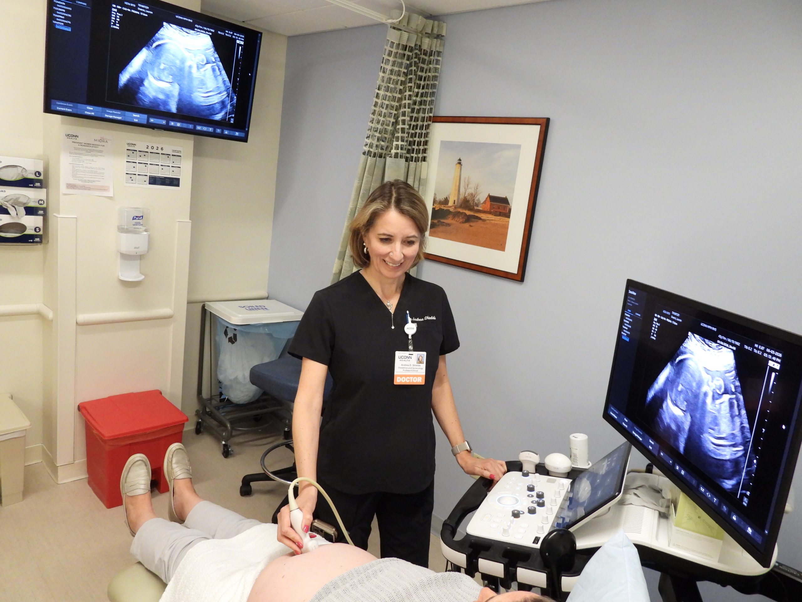
Courtois, JAX’s associate director of the Single Cell Biology group, in collaboration with Luciano, UConn School of Medicine Associate Professor of Obstetrics & Gynecology, recently completed an important study to develop a comprehensive cell atlas of the disease based on lesions obtained from 14 individuals who had treatment for endometriosis at UConn Health’s Center of Excellence in Minimally Invasive Gynecology.
“Single-cell analysis of endometriosis reveals a coordinated transcriptional program driving immunotolerance and angiogenesis across eutopic and ectopic tissues,” a paper published in Nature Cell Biology, includes a thorough comparison of healthy endometrium tissue and ectopic (outside their normal site) lesions. The data also describes the endometriosis microenvironment and the conditions that allow the lesions to form and grow in what should be unhospitable regions.
“The study builds a robust foundation for a better understanding of endometriosis and how it grows,” says Luciano of OB/GYN at UConn Health’s Women’s Center. “It’s exciting progress that we hope leads to earlier diagnosis and the ability to specifically target these abnormal cells for better treatments.”
Growth and immune escape
The research team, which also included first author and Predoctoral Associate Yuliana Tan, Ph.D., and Associate Professor and Director, Single Cell Biology Paul Robson, Ph.D., who is also a faculty member in UConn’s Department of Genetics and Genome Sciences, worked with tissues from individuals who had lesion removal at UConn Health for relief of symptoms. All were also receiving hormone therapy, the most frequent endometriosis management strategy. Not surprisingly, given that lesions are described as endometrial-like tissues growing in the wrong place, the cellular composition of the lesions in the peritoneum were quite similar to that of the normal endometrium. On the other hand, ovarian lesions had extensive differences in both composition and gene expression from the peritoneal ones. So while both ovary and peritoneum are receptive to the formation of lesions, they represent different environments and lead to important cellular and molecular differences between the two sites. The finding indicates that site-specific therapeutic design may be necessary to develop more effective treatments.
Another aspect of endometriosis is that, like cancer, the lesions represent abnormal growth that would typically be eliminated by immune surveillance. The researchers therefore investigated the immune cells in the peritoneal lesion microenvironment to see why they do not eliminate the abnormal lesion cells. They found that two important immune cell types, macrophages and dendritic cells, contribute to conditions that promote immune inhibition and the promotion of immunosurveillance escape. Their specific characteristics are similar to those associated with fetal tolerance during pregnancy, suggesting that endometriosis hijacks a necessary, naturally occurring immune process to allow for lesion formation and persistence.
Improving endometriosis knowledge and care
The paper details other aspects of both normal endometrium and ectopic lesions, including properties of vascularization and the drivers of regeneration in endometrium and, perhaps, the formation of lesions in endometriosis. Of particular interest were key differences in the vascularization (blood vessel formation) of peritoneal versus ovarian lesions, further emphasizing the site-specific nature of endometriosis. Also of note was the identification of a previously uncharacterized population of epithelial cells that may be progenitor cells for both endometrium and lesion formation, but more work is needed to define their precise role.
“Single cell analyses and hyperplexed antibody-based imaging techniques offer powerful insights into the complexity of the endometriosis microenvironment,” says Courtois. “Understanding this complexity will be key for developing the new, efficient diagnostic and therapeutic tools that are so badly needed.”
Overall, the data captures a full description of endometrium and lesions, laying a strong foundation for understanding the vital cellular players and molecular dynamics of the disease. The data represents an important step forward for research into endometriosis and provides essential information for future therapeutics and diagnostics that can provide relief for those with this under-investigated disease.