UConn researchers are working with college athletes to test a new device that can quickly assess concussions and other traumatic brain injuries.
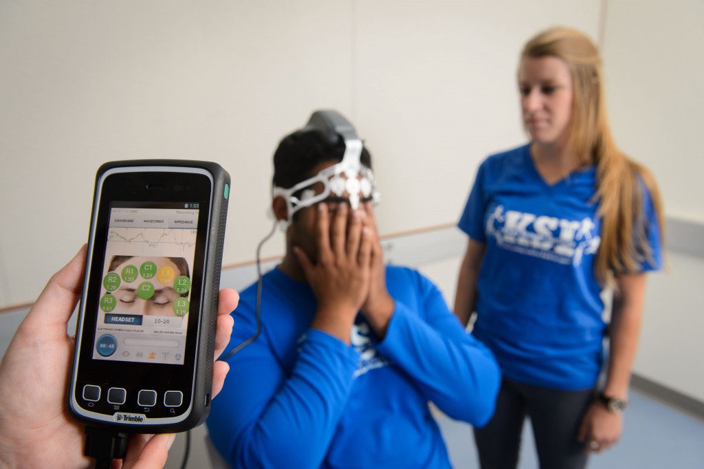
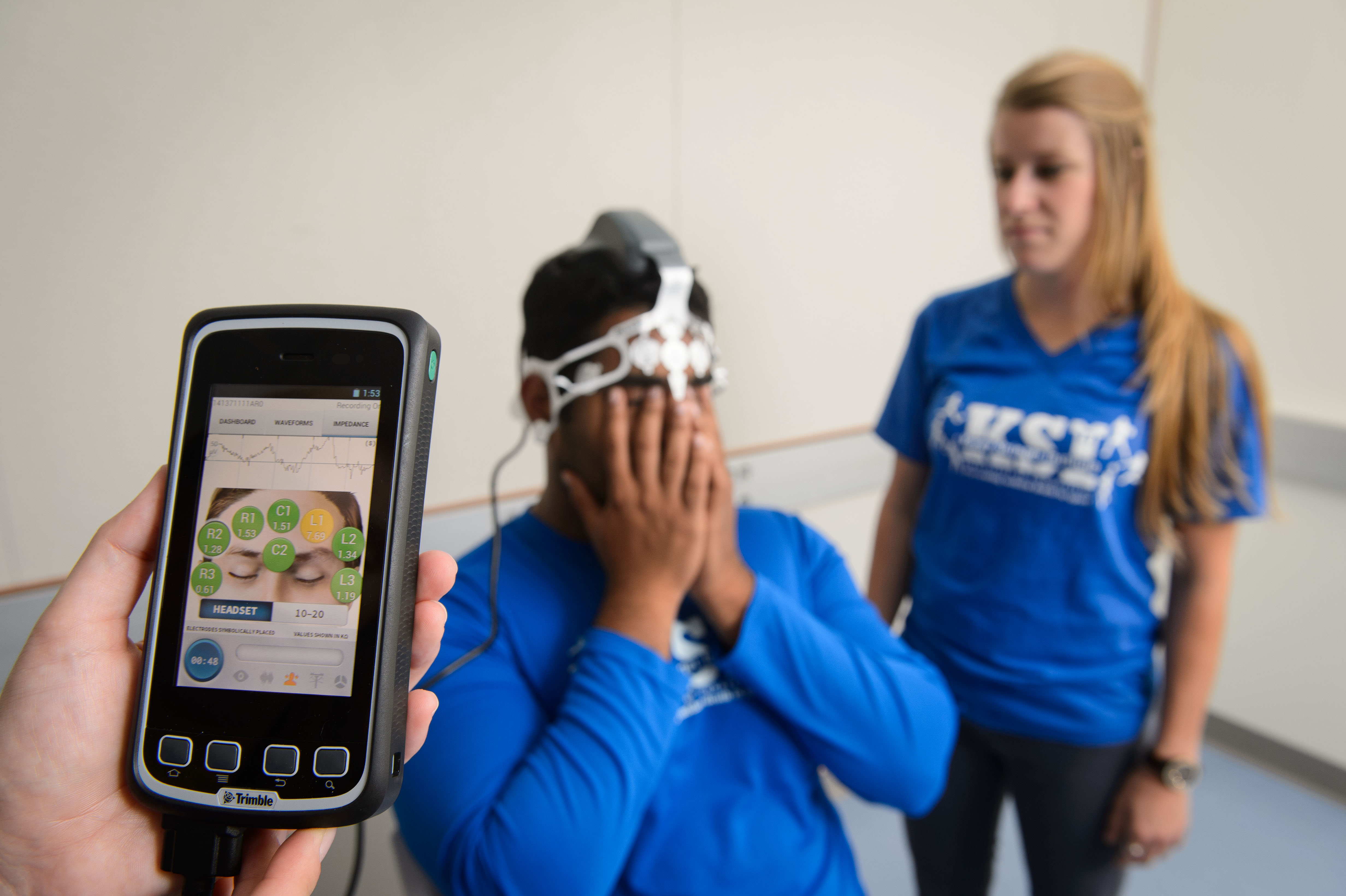
The device, developed by Bethesda, Md.-based medical neuro-technology company BrainScope Co. Inc., is a handheld instrument that can help clinicians identify traumatic brain injury (TBI) at the time and place of injury.
“BrainScope approached the Korey Stringer Institute because of our extensive background in conducting research studies,” says UConn graduate student Samantha Scarneo, director of youth sports safety at the Korey Stringer Institute. The concussion study “aligns with KSI’s overall mission to prevent sudden death in sports and overall safety in all levels of sport,” she adds.
The device, which is not yet available commercially, is about the size of a smartphone. Placed on a patient’s head, it measures a patient’s electroencephalograph (EEG), or brainwaves, to gauge brain function after head injury.
The non-radiation-emitting instrument was developed for military use in war zones, and is being adapted for athletes who sustain a TBI while playing contact sports. Within 10 minutes, the device can help medical personnel determine whether it’s safe for a player who’s had a head injury to return to the athletic field.
TBIs include structural injuries such as a bruise or bleeding in the brain and what experts call a “milder form” of TBI, concussions, caused by a bump or jolt to the head. These injuries can alter brain function yet be difficult to detect, as they don’t always cause immediate symptoms.
UConn, one of nine universities participating in the nationwide study, offers an added benefit in the form of state-of-the-art MRI equipment at the University’s new Brain Imaging Research Center, and scientific staff to analyze and make sense of the diagnostic and medical imaging data generated by the study.
Peter Molfese, assistant research professor and director of operations at UConn’s Brain Imaging Research Center, notes that MRI works by manipulating magnetic fields of hydrogen molecules in the body to identify the structure and function of different brain tissues.
After a concussion or other traumatic brain injury, Molfese says, there is a reduction in both gray and white matter in the brain. “Less brain means that functions that were otherwise handled by particular areas need to be remapped to other areas. However, the brain’s ability to remap function is not perfect, and various levels of head injury can leave people with permanent brain damage that can hinder their abilities later on.”
Research efforts to accurately and quickly evaluate TBIs have intensified in the U.S. because of the toll they have had on the U.S. military (300,000 cases since 2000, according to the Department of Defense), and high-profile lawsuits brought against the National Football League by former players for its handling of concussions. In addition, a recent Centers for Disease Control and Prevention report found that 2.5 million emergency department visits, hospitalizations, or deaths were associated with TBIs.
The UConn researchers are now working with both UConn club sports teams and Eastern Connecticut State University varsity and club sports teams to enroll athletes for the study. They aim to complete data collection in December or by the end of the spring sports season in 2016, depending on the number of athletes who sustain concussions. The study will use the Brain Imaging Research Center’s fMRI machine for all of its MRIs, on both injured and non-injured participants.
The interdisciplinary team working on the study includes Douglas Casa, head of the Kinesiology Department’s Korey Stringer Institute, and Jeffrey Kinsella-Shaw, director of the physical therapy doctoral program, in addition to Scarneo and Molfese.


