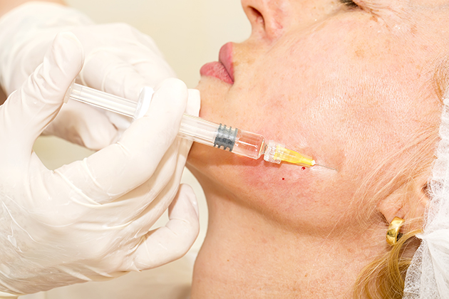
An off-label use of Botox has been found to be effective in relieving temporomandibular joint (TMJ) dysfunction syndrome and associated myofascial pain. But an oral radiologist at UConn Health is concerned that the short-term benefit may come at a price.
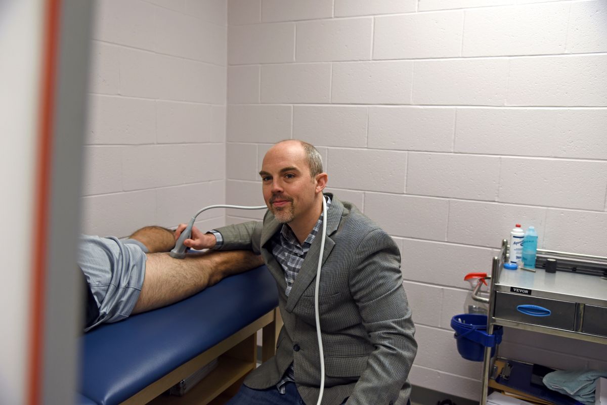
Dr. Aditya Tadinada, assistant professor of oral and maxillofacial radiology at the UConn School of Dental Medicine, was involved in a pilot study that finds evidence of bone density loss in the facial skeleton associated with botox injections – enough evidence that the National Institutes of Health is now funding a five-year, $4 million, multi-site clinical trial. Tadinada is the radiologist on the study, collaborating with Dr. Karen Raphael from New York University (NYU).
“Botulinum toxin is injected into inflamed muscles, and what really happens is these muscles undergo paralysis,” Tadinada says. “Over prolonged use, they cause a constant unloading of the temporomandibular joint, leading to a loss in the density of the bone. That could potentially lead to pathological fractures, and also sometimes to necrosis or flaccidity of the muscle itself.”
Think of the TMJ as the hinge at the back of the jaw that enables us to open and close our mouth. Myofascial pain syndrome is a chronic condition involving pain and inflammation in a single muscle or muscle group that can trigger discomfort in other areas of the body.
NYU and some clinics in the Los Angeles area have been identified as intake centers for the study. The researchers are considering additional locations to expand the scope and outreach.
The study primarily will use a new low-dose 3-D imaging modality known as cone beam computed tomography (cone beam CT) to evaluate the changes in bone density. Tadinada expects to start reading images early next year. The researchers hope to recruit at least 100 patients across the country.
“While the patients are getting temporary relief from the pain – and those patients really need help, because it’s a terrible amount of pain – we do not know the long-term consequences of this off-label use of the drug,” Tadinada says. “We don’t have any studies that show us. The company that makes Botox is not interested, because that’s not the primary reason they’re selling it.”
Rare breed
There are fewer than 10 oral and maxillofacial radiology residency programs in the United States, and UConn’s, which goes back to 1973, is the oldest.
“Our radiology program is world renowned,” says Dr. Alan Lurie, chair of the Division of Oral and Maxillofacial Diagnostic Sciences at the UConn School of Dental Medicine. “We have people running dental radiology departments all over the world.”
Tadinada is a product of that residency program. Now that he’s on the faculty, he and Lurie are involved in the Alliance for Radiation Safety in Pediatric Imaging’s “Image Gently” campaign to promote the use of as little radiation as possible when taking dental X-rays.
“We are gatekeepers for responsible imaging, especially in pediatrics,” Tadinada says. “We’ve revised our protocols to use the lowest dose of radiation possible. One way we accomplish this is by narrowing the field of vision. Our training in both dentistry and radiology enables us to do this.”
Oral radiologists are part of an elite group, and this year Tadinada joined an even more elite group: The American Academy of Oral and Maxillofacial Radiology awarded him with its highest research honor for junior faculty, the William H. Rollins Award.
William Rollins was the first radiation health physicist, and the first to warn that X-rays can be dangerous. The award goes to a junior faculty member who has demonstrated a significant body of research work on imaging sciences, and is not given every year.
Making dental imaging safer
Tadinada’s investigation of Botox is just one example of the research he’s doing to improve dental patient safety. Another has to do with exposure to radiation during dental imaging, and he may be closing in on a way to eliminate it. His lab is working on an alternative imaging method known as optical coherence tomography.

With this approach the wavelength is altered, so rather than penetrate tissue, the light is reflected back. It’s similar in concept to ultrasound, but uses light instead of sound.
“We’re basically throwing light photons at the patient, and we wait to see how they come back and at what point they come back, and based on this, we give each of these signals a coordinate, and we’re able to make an image out of it,” Tadinada says. “UConn is one of the very few centers in the whole country that is even looking at using this in the mouth.”
He already has been able to capture some tooth images this way. The ability to take images without the use of ionizing radiation would open new doors in image-assisted dentistry.
“I think where this will end up is similar to cardiac imaging, where they keep that live feed so they can see where they’re going – which would be amazing for the practitioner, no matter what the procedure is,” Tadinada says. “With the heart it’s worth the exposure risk because of the criticality of the organ, but not with the mouth. My goal with this is, if there is not ionizing radiation, to leave it on, so the dentist is able to see on the screen where he is going with the cavity prep, for example.”
Follow UConn Health on Facebook, Twitter and YouTube.