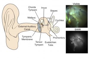
This diagram shows the structures of the middle ear, along with examples of the kinds of images provided by today’s conventional visible-light otoscopes (top) and by the newly developed short-wave infrared (SWIR) otoscope. The new otoscope can probe deeper to provide clearer indications of the presence of fluid which can indicate an infection. (Courtesy of the authors and PNAS)
PNAS-single column
This diagram shows the structures of the middle ear, along with examples of the kinds of images provided by today’s conventional visible-light otoscopes (top) and by the newly developed short-wave infrared (SWIR) otoscope. The new otoscope can probe deeper to provide clearer indications of the presence of fluid which can indicate an infection. (Courtesy of the authors and PNAS)


