UConn School of Dental Medicine’s advanced imaging capabilities and research pursuits are now being showcased at Yale University’s medical library exhibit until Nov. 3.
The exhibit “New Lives for Old Specimens” unveils new ways collections of historical anatomy and pathology specimens are being used for new research and medical education initiatives.

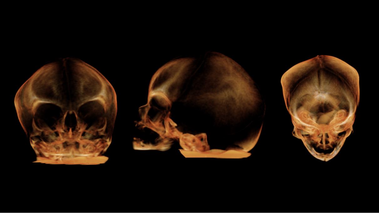
For high-tech imaging analysis of the exhibit’s showcased historical specimens of fetal skulls from Quinnipiac’s Kier/Conlogue collection, researchers turned to UConn’s dental school for its expertise.
A research team led by UConn dental school’s Dr. Alan Lurie used the most advanced Cone Beam CT imaging on the skulls. The research project has taken hundreds of volunteer hours since 2015. In addition to Lurie, the UConn research team includes Dr. Sonya Kalim, Dr. Aditya Tadinada and Andrew Emery, a third-year dental student.
Cone Beam CT imaging is the most rapidly growing advanced imaging for dental patients needing dental implants or root canal care and for ENT patients, according to Lurie. This advanced imaging allowed researchers to precisely measure in millimeters areas of the skulls, count present teeth and stage the teeth’s development stage. Currently, dental and medical textbooks report that fetal teeth start to develop as early as six to eight weeks in the womb starting with primary teeth.
The Cone Beam CT imaging technology allows for a 360 degree panoramic view of the inside and outside of each skull in the most highly-specialized 3D high-resolution, and advanced software imaging helps create high-tech skull and facial bone imaging reconstructions.
The team’s imaging and preliminary research findings now on display at Yale indicate potential facial, dental and cranial developmental abnormalities of the skulls, which may help gain further insights into the human face’s developmental process at the earliest stages of life.

Dr. Aditya Tadinada; and is led by Dr. Alan Lurie (Photo: UConn School of Dental Medicine).
In this ongoing skull imaging research project, the UConn dental imaging research team hopes to pinpoint the best ways to image the human face and skull, and improve scientific knowledge of how the human face actually develops.
“The human face’s developmental process may be different than how our textbooks currently teach us,” stresses Lurie. “We look forward to further research analysis and publishing our team’s research findings.”
The exhibit is a large collection of projects on display involving historical specimens ranging from brain tumors to skulls. The exhibit is a collaboration between multiple curators from inside and outside of Yale School of Medicine, and medical students, faculty and research teams hailing from UConn School of Dental Medicine, Quinnipiac and others from Ireland and Canada.
The exhibit is open to the public May 25 to Nov. 3 at Yale’s Harvey Cushing/John Hay Whitney Medical Library located in the Cushing Rotunda at 333 Cedar Street in New Haven. For more information, visit: here.