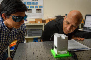
A portable holographic field microscope developed by UConn optical engineers could provide medical professionals with a fast and reliable new tool for the identification of diseased cells and other biological specimens.
The device, featured in a recent paper published by Applied Optics, uses the latest in digital camera sensor technology, advanced optical engineering, computational algorithms, and statistical analysis to provide rapid automated identification of diseased cells.
One potential field application for the microscope is helping medical workers identify patients with malaria in remote areas of Africa and Asia where the disease is endemic.
Quick and accurate detection of malaria is critical when it comes to treating patients and preventing outbreaks of the mosquito-borne disease, which infected more than 200 million people worldwide in 2015, according to the Centers for Disease Control. Laboratory analysis of a blood sample remains the gold standard for confirming a malaria diagnosis. Yet access to trained technicians and necessary equipment can be difficult and unreliable in those regions.


