While mammograms have been proven necessary screening for early signs of breast cancer, women typically don’t look forward to the uncomfortable annual visit. But now, new low dose mammography technology available at UConn Health captures clearer images with less time in compression and half the dose of radiation.
In the past year, the Beekley Imaging Center at UConn Health has converted standard digital mammography to low dose mammography, which uses synthetic 2D mammogram technology, to continue providing high-quality breast imaging services.
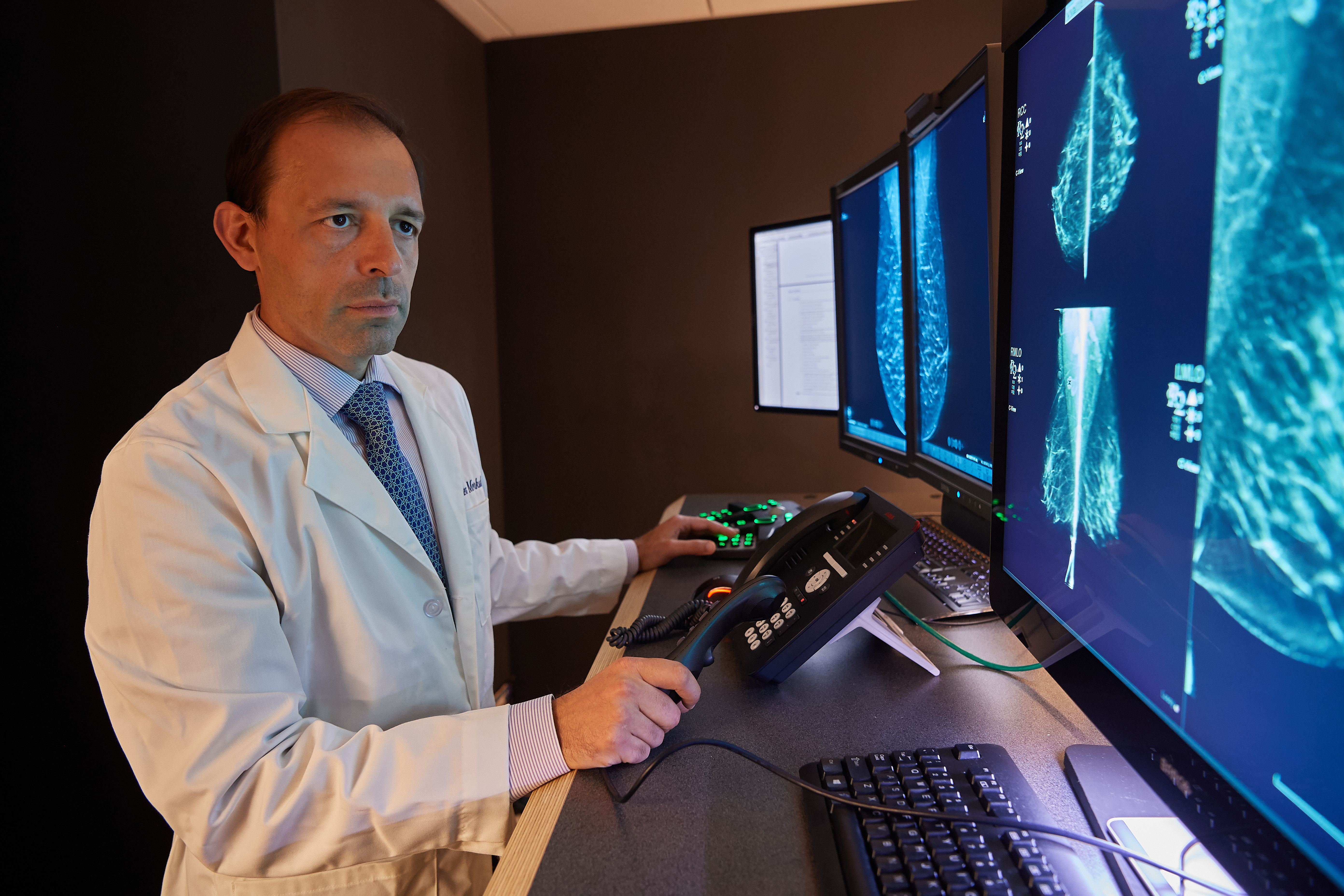
“UConn Health is proud to be on the forefront of innovation in women’s imaging, offering the latest FDA-approved technology with low dose mammography to diagnose breast cancer earlier,” says Dr. Alex Merkulov, section head for Women’s Imaging at UConn Health.
Breast cancer is the second most common cancer among women in the United States, and early detection reduces the risk of death by at least 25 to 30%, according to the American Cancer Society. Based on current guidelines, UConn Health recommends that women should begin having mammograms yearly at age 40, or earlier if they’re at high risk.
“Low dose mammography is a huge leap forward in detecting breast cancer early,” says Merkulov.
The technology used for this implementation, made by Hologic and called Low Dose 3-D Mammography with C-View Software, instantly generates a 2D image directly from the tomosynthesis data. Each breast is compressed once, and a machine takes many low dose X-ray images as it moves in an 11-degree arc over the breast. A computer then puts the images together into a series of thin slices, allowing doctors to see breast tissues more clearly in three dimensions.
The technology lowers the patient’s dose of radiation and reduces compression and exam time compared to traditional mammograms. The safety and efficacy of the C-View 2D screening exam have been validated through multiple clinical evaluations.
Early detection is the best defense against breast cancer, and the 3D breast images from this technology help doctors detect breast cancer earlier than previously possible, Merkulov says.
Since 2007, breast cancer death rates have been steady in women younger than 50, but have continued to decrease in older women. From 2013 to 2017, the death rate decreased by 1.3% per year, according to data from the American Cancer Society.
These decreases are believed to be the result of breast cancer being found earlier through screening and increased awareness, as well as better treatments.
Many studies have found that 3D mammography appears to lower the chance of being called back for follow-up testing. It also appears to find more breast cancers, and several studies have shown it can be helpful in women with dense breast tissue. The technology provides peace of mind, detecting abnormalities that are missed by traditional mammograms, delivering more detail and accuracy.
Patients can now schedule a screening mammography without a physician referral online through their MyChart account or by calling 860-679-3634.