Every time you walk, run, or pick up a cup of coffee, your joints are at work. For millions of people with osteoarthritis, a degenerative joint disease, these simple movements are incredibly painful.
Osteoarthritis affects more than 66 million Americans and costs approximately $303 billion a year to manage the disease.
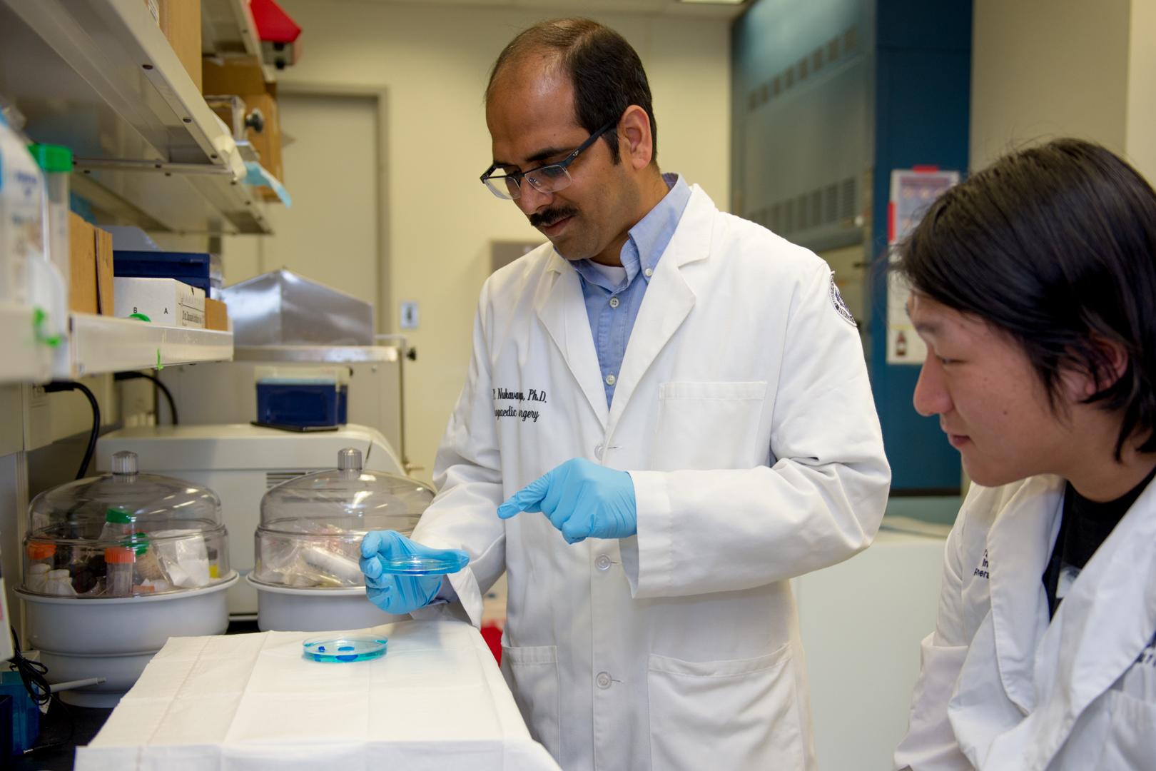
Syam Nukavarapu, an associate professor of biomedical engineering at the University of Connecticut, is developing novel tissue engineering technologies that could radically improve treatment for this condition. He also holds joint appointments with materials science and engineering, and orthopedic surgery at UConn’s Schools of Medicine and Dental Medicine.
There are currently two primary osteoarthritis treatments: microfracture surgery and tissue replacement using autografts (tissue from oneself) or allografts (tissue from another). Microfracture surgery creates tiny fractures in underlying bone, spurring the cells to create new cartilage. This often creates new tissue that is mechanically inferior and can cause further degeneration over time. Autografts or allografts replace damaged or diseased tissue with donor tissue, which can be a problem if donors are not readily available. This method also carries concerns of disease transmission and failure of the transplant over time. Neither treatment is ideal or guaranteed.
Nukavarapu’s tissue engineering approach to repairing joint tissue is a promising avenue for the development of new treatments, which involves fabricating synthetic bone-cartilage or “osteochondral” grafts that can be implanted within the body to promote new tissue formation and repair.
Scientists have found ways to generate various tissue types like skin or bone using tissue engineering principles. But this becomes much more challenging when dealing with complex tissues such as joints that consist of bones covered by a layer of cartilage – two dissimilar tissues connected together, which are referred to as “tissue-tissue” interfaces.
Designing a graft to support bone and cartilage regeneration along with an interface is an immense engineering challenge.
“In complex tissue repair, the biggest problem is not just regeneration, but the tissue-tissue integration with an interface,” Nukavarapu says.
Despite this challenge, Nukavarapu and his lab (Tissue Engineering Science and Technology Lab, TEST Lab) are having success. They recently published research on a gradient graft platform for interfacial tissue engineering in the December 2020 issue of Biomedical Materials. Deborah Dorcemus, an alum of Nukavarapu’s lab, and Hyun Kim, a current Ph.D. student, are co-authors. The novelty of this approach led to a successful patent as well, paving the way for future applications.
With this award, TEST Lab will design and develop scaffold platforms using biodegradable materials with desired configurations and geometries. In this setup, local biomaterial stiffness and microenvironments can be manipulated to precisely control the differentiation of stem cells into specific cell types.
The gradient structure of Nukavarapu’s engineering graft gradually transitions from a more porous cartilage layer to a dense bone layer. This appears to be a more effective approach than previous designs, because it provides structural flexibility and mechanical integration while accommodating two dissimilar tissue types in a single graft structure. The cartilage layer is filled with a gel to imitate the structural and physical properties of the cartilage tissue, which is naturally flexible and soft.
“We provide an ideal mechanical environment in terms of the local micro-structure and properties for cartilage repair,” Nukavarapu says.
The other key feature of this scaffold is the ability to spatially provide growth factor substances that recruit and/or stimulate cells – in this case, cells that give rise to cartilage and bone tissue.
Nukavarapu incorporated bone-specific growth factors throughout the gradient structure, which will give rise to bone, and a chondrogenic growth factor within the gel, which will give rise to the cartilaginous layer. So far, the results showed that the growth factors were properly localized in the structure resulting in a scaffold with a spatial growth factor profile needed to approach complex tissue engineering, such as for joints.
While this technology is promising, Nukavarapu’s lab continues to develop new biomaterial-centered strategies for tissue engineering. His laboratory has demonstrated the role of biomaterial stiffness and topographical features (micro- and nano-sized) that can regulate cell behavior.
This research fueled his new NIH-funded grant, where Nukavarapu and his lab are looking at how to use physical and structural cues in tissue engineering strategies to control cellular function and regeneration.
With this award, Nukavarapu and his lab will design and develop scaffold platforms using biodegradable materials with desired configurations and geometries. In this setup, local biomaterial stiffness and microenvironments can be manipulated to precisely control the differentiation of stem cells into specific cell types.
The goal of this project is to implement geometric and physical cues in a three-dimensional setting to design synthetic grafts for complex tissue engineering. Specifically, bone, cartilage, and bone-cartilage interface development.
These efforts will utilize additive manufacturing methods to create scaffold structures with precise microstructure, stiffness, and environment. The reproducibility of manufacturing methods such as bio-printing will allow Nukavarapu and his team to uncover new ground in the area of interface engineering.
This project will push the frontier of tissue engineering even more by developing new image modalities to correlate biomaterial stiffness and geometry with regenerated tissue quality and its function. Achieving a stem cell niche – the conditions necessary to stimulate stem cells to produce a specific tissue type – could provide manifold improvements in osteoarthritis treatment.
“We’re trying to take tissue engineering to the next level,” Nukavarapu says. “If we succeed with this, we can implement a biomaterial-centered paradigm for tissue engineering.”
In this effort, Nukavarapu is collaborating with UConn Health researchers Sangamesh Kumbar and Vinayak Sathe.
Ultimately, the novel graft system with the support of the new NIH funding could advance the development of new osteoarthritis treatments, which would make that walk or run, perhaps to pick up your favorite coffee, that much easier.
Part of this work is supported by the Office of the Vice President for Research (OVPR) SPARK Technology Commercialization Fund and BioInnovation CT.