Leading the way in breast-conserving surgery, UConn Health is the first hospital in New England to offer the surgical navigation system by EnVisio.
Approximately 192,200 women in the United States are diagnosed annually with invasive breast cancer.
Breast-conserving surgery (BCS) may be used as part of a treatment plan for breast cancer. It is sometimes called a lumpectomy or a partial mastectomy. During BCS, the cancer lump and some breast tissue around the lump are removed.
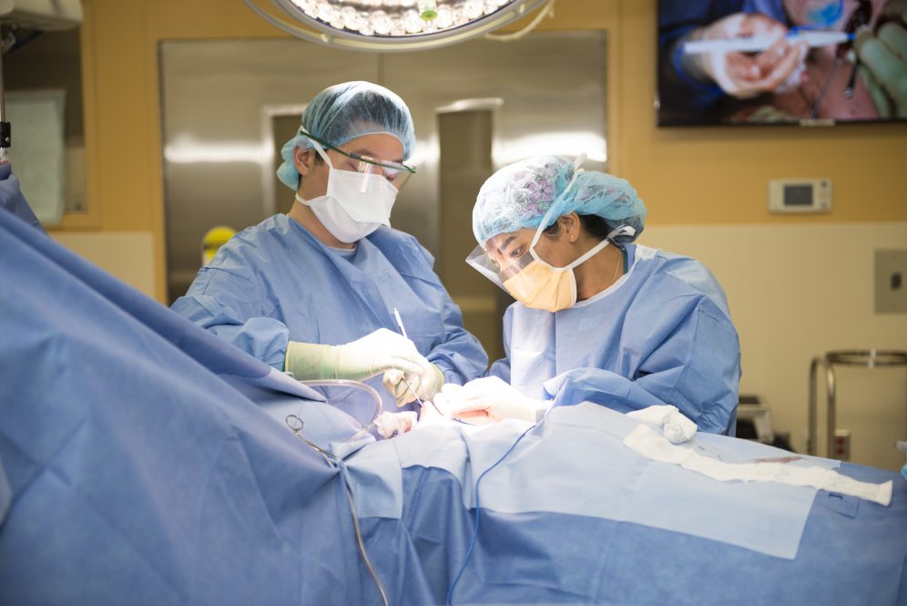
The EnVisio™ Navigation System is currently being used by Dr. Christina Stevenson, associate professor of surgery at UConn Health, and Dr. Dana Scott, assistant professor, department of Obstetrics and Gynecology, at UConn Health.
The 3D surgical navigation system provides the surgeon a precise pathway to the targeted tissue where the Department of Radiology team has already placed the SmartClip™. This helps localize lesions in the breast that the surgeon cannot feel.
The SmartClip™ is used by surgeons with the EnVisio™ Navigation System to identify the malignancy location and wirelessly navigate to it in real-time 3-D. The system can uniquely identify three different SmartClip™ Soft Tissue Markers – allowing physicians to effectively mark difficult lesions and navigate distances, depths, and directions.
Normally on the day of surgery, a radiologist will insert a hook wire through the skin down to the biopsy clip. This hook wire protrudes from the skin and provides a visible road map to the lesion. The surgeon makes an incision in the skin and follows along the wire to the target to remove the marker, wire, and lesion. By eliminating the hook wire on the day of surgery, the new marker promises to streamline scheduling and reduce patient stress, pain, and costs.
“Patients are already going through a stressful time so to be able to alleviate some of that anxiety by having the SmartClip™ technology is important for our patients,” says Stevenson.
With this new technology, a specialized clip is placed by the radiologist any time ahead of the surgery which streamlines the day of surgery. The clip is less uncomfortable than having the wire that protrudes through the skin. It is also FDA approved to stay in the patient indefinitely, making it easier if the need for a future procedure arises.
The marker provides the distance, depth, and direction information of its location to an electronic pen-like device that the surgeon uses during tumor removal.
The technology allows the surgeon to target the mass in 3 dimensions so it can be removed without having to take more tissue than is needed. Accurate localization of the SmartClip also aids the surgeon to remove an adequate margin of normal tissue surrounding a tumor.
Stevenson had been researching the technology with several companies and what made this one more attractive to use as a platform at UConn Health was that there was no radiation involved during the procedure. Some of the competitive technologies had limits to how long the clip could stay in, but this one is FDA approved to stay in indefinitely.
For more information visit the UConn Health Carole and Ray Neag Comprehensive Cancer Center.