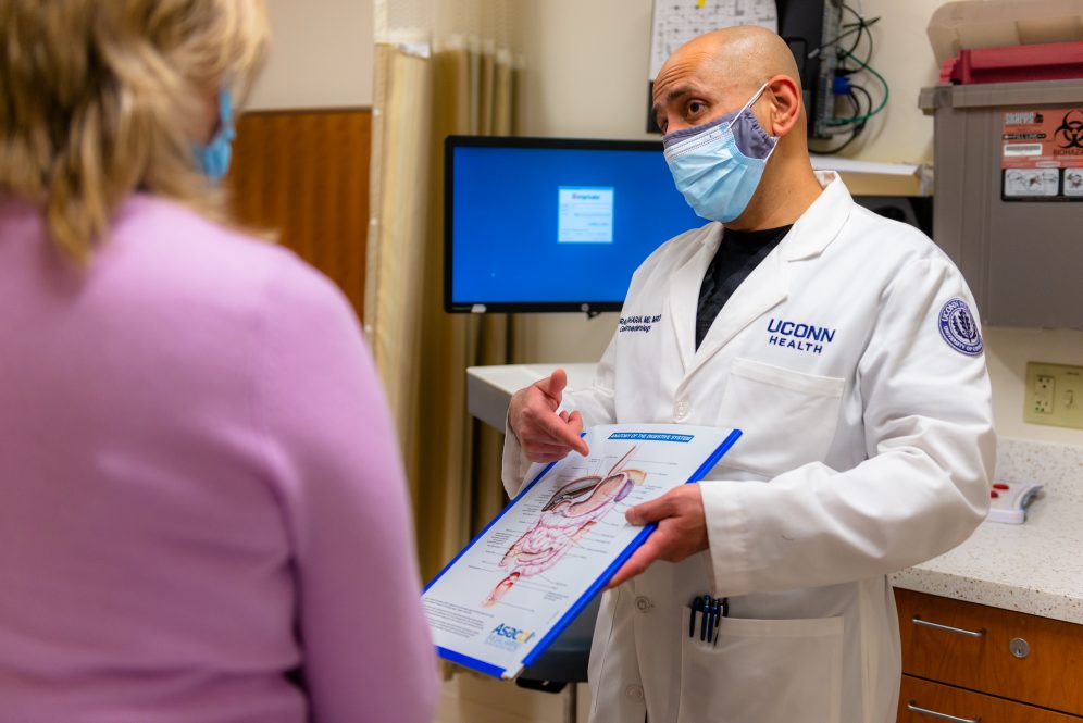
The use of endoscopic ultrasound (EUS) takes a steady hand, particularly when venturing on the outer edge of the EUS frontier.
That’s where you’ll find Dr. Murali Dharan: at the forefront of UConn Health’s advanced endoscopic techniques.
Most notably, Dharan recently performed the first successful endoscopic ultrasound-guided gallbladder drainage procedure in central Connecticut. It’s a new option for patients who otherwise might be at high risk of complication from gallbladder removal surgery or might need an external catheter to drain the gallbladder outside the body.
Instead of penetrating the abdominal wall to insert a tube to drain the gallbladder or drain inflammatory cysts from the pancreas, Dharan uses endoscopic ultrasound and steady hand-eye coordination to place what’s known as a lumen-apposing metal stent, which enables internal drainage, no external catheter needed.
“It’s a special stent, shaped almost like a dog bone, and under EUS guidance, you are potentially able to look at any structure that is close to the stomach or intestine,” Dharan says. “Be it the gallbladder, be it the bile duct, be it a big cyst, you are able to visualize it. So once you see that structure and you are able to line it up, you make a small puncture and put the stent in.”
Dharan arrived in August of 2019 with a mission of growing UConn Health’s advanced endoscopic capabilities. Another such procedure is the cholangioscopy, which he and Dr. John Birk, chief of UConn Health’s Division of Gastroenterology and Hepatology, have just started offering.
“With cholangioscopy, you pass a very thin camera right into the bile duct itself, so you visualize the bile duct as you would visualize the stomach or intestine with an endoscope,” Dharan explains. “For complex stone disease or complex strictures of the bile duct including cancer, where you cannot get an easy diagnosis, we are now able to directly visualize the lesion and perform a biopsy to confirm what is going on or offer direct therapy such as breaking the stones with ultrasonic pulses.”
When a patient needs both an endoscopy and a biopsy, of the liver for example, Dharan can do both endoscopy and EUS-guided liver biopsy during the same procedure, while the patient is sedated, sparing him or her a needle biopsy through the rib cage.
The charge to elevate advanced endoscopy services drew Dharan to UConn Health, and so did the appeal of working with trainees. He recalls encountering residents and fellows while practicing at Saint Francis Hospital and Medical Center.
“The more I interacted with them, the more I realized I wanted to go into academics,” Dharan says. “At the UConn School of Medicine, I’m focusing on the slightly advanced issues of endoscopy. I work more with the senior GI fellows to highlight the finer aspects of the advanced endoscopy techniques.”
Whether he’s talking to future or current gastroenterologists, Dharan is quick to emphasize the untapped capabilities of endoscopic ultrasound.
“There are a lot of applications, some of which I’ve already brought to UConn,” he says. “But I think the more people understand what EUS has to offer, the more we can develop the service to realize its full potential.”
This story originally appeared in the Spring 2021 issue of UConn Health Journal.