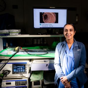
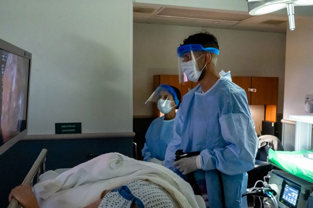
Gastroenterologists and cancer prevention specialists at UConn Health emphasize the importance of colonoscopies as an effective tool to prevent colorectal cancer.
Now they’re using the most advanced technology they’ve ever had to do so.
W
e can better identify abnormalities in the intestinal lining, and more subtle changes, and hopefully therefore pickup up pathology at an earlier time. — Dr. John Birk

UConn Health is an early adopter of an endoscopy system that provides a new level of clarity to physicians examining the gastrointestinal tract.
“Colonoscopy has been widely adopted as a screening mechanism for colon cancer, and been shown to be effective in lowering your risk of mortality from colon cancer,” says Dr. John Birk, chief of UConn Health’s Division of Gastroenterology and Hepatology. “And we’re getting even better at it. With our new instruments, we can better identify abnormalities in the intestinal lining, and more subtle changes, and hopefully therefore pickup up pathology at an earlier time.”
It’s a simple formula that applies to many illnesses: Find it soon enough to address before it becomes problematic. Colonoscopies enable physicians to locate, identify, and remove either cancerous lesions before they can spread, or other growths before they have a chance to become cancerous.
The applications go beyond colon cancer screening. Endoscopy helps diagnose and treat acid reflux, ulcers, Crohn’s disease, and celiac disease, some of which potentially can lead to other cancers.
The new endoscopy system includes a range of new technologies that enhance image color and texture, improve visibility of deep blood vessels and bleeding points, and make darker areas of the endoscopic image easier to see.
It all adds up to clearer, better-defined imaging that enables physicians to instantaneously identify abnormalities and help inform next steps during the procedure.
“Examples of enhanced imaging helping us in upper endoscopy are identifying Barrett’s esophagus more easily, like someone with chronic anemia who may have minor bleeding, and is not bleeding actively at the moment,” Birk says.

Barrett’s esophagus, which is when acid reflux causes damage to the esophagus, can be a precursor to esophageal cancer. Another condition that calls for screening is atrophic gastritis, or chronic inflammation of the stomach lining’s mucous membrane, which can be a precursor to gastric cancer.
“This new enhanced imaging will help identify abnormalities in these precursor lesions too, and it actually may help even more [than with colorectal cancer],” Birk says.
Another advantage is the endoscopes themselves are lighter and easier to handle, and the tools used for upper GI procedures are thinner, causing less of a disturbance to the patient’s throat.
The technology, which is from Olympus, also could lead to growing acceptance the optical biopsy, a method of using imaging to identify whether a polyp is more serious or benign without having to remove the tissue and send to the pathology lab.
The American Cancer Society’s guidelines are for people considered to be at average risk start to regular colorectal cancer screening at age 45. Because factors in determining risk can include family history, medical history, lifestyle, and demographics, UConn Health’s Colon Cancer Prevention Program works with each patient to create an individualized plan for recommended timing, method, and frequency of screenings.
To schedule an appointment or learn more, call the Colon Cancer Prevention Program at 860-679-4567, or the Division of Gastroenterology and Hepatology at 860-679-3238.


