When a pathogen enters the body, the immune system responds by mounting an attack against it.
Seems simple. But this description doesn’t account for why some people get much sicker than others from the same exposure.
Jianbin Ruan, assistant professor of immunology at UConn School of Medicine, leads a lab focused on discovering the molecular mechanisms at work in immune signaling to provide answers to this question and pathways for more effective treatments and vaccines.
Ruan’s Ph.D. work at the University of Science and Technology of China focused on epigenetics – not immunology.
Humans share 99.9% of their DNA. Yet, people have vastly different health outcomes. Epigenetics, the study of gene expression changes that occur without changes to the underlying DNA, helps us understand why this happens.
As Ruan was preparing to graduate, he was seeing more and more scientific literature about unexpected engagement of proteins in immune signaling. Ruan pivoted during his postdoctoral fellowship at Harvard Medical School where he studied how immune signaling mediated responses to infection.
“In the big picture, epigenetics and immunology are quite similar,” Ruan says. “Everybody has the same genes, but they can have different shapes, different looks, and different behaviors. A lot of that is influenced by epigenetics. The immune system works in a similar way — people may share the same immune genes, yet respond to infections differently.”
Ruan’s lab at UConn Health focuses on three areas of research: non-canonical inflammasome signaling, gasdermin-mediated programmed cell death, and pore-forming proteins/toxins.
“In my lab, the most exciting part, although it’s very basic, it’s also the most fundamental part –understanding how our immune system works at the molecular level, especially immune signaling,” Ruan says. “The ultimate goal of all the projects is to try to understand the basic mechanisms of how the immune system works and how bacteria manipulate our signaling and to try to provide some ideas for clinical translation of those studies.”
Canonical inflammasomes, a type of protein complex that has been studied extensively, rely on sensor proteins to detect infection or cellular damage. This signals the cells to release cytokines (signaling proteins that help coordinate immune responses) and induces inflammatory cell death to eliminate infected cells.
Non-canonical inflammasomes, by contrast, are not well studied. These proteins sense lipopolysaccharide (LPS), a major component of the outer membrane of Gram-negative bacteria, in the cytosol (the liquid inside cells). Recognition of cytosolic LPS triggers inflammatory cell death, a key driver of sepsis.
Previous studies found that LPS with six lipid chains trigger non-canonical inflammasomes. But different bacteria have different LPS with more or fewer lipid chains. LPS with only five lipid chains, for example, do not activate the inflammasomes.
A recent study Ruan published in Science Advances demonstrated that the number of lipid chains in LPS dictates activation of the non-canonical inflammasome.
“We study how, in response to upstream secondary activation of what’s called the ‘executioner protein’, it causes immediate cell death and how this kind of cell death can affect our immune system,” Ruan says. “Gasdermin is responsible for this process.”
Recently, Ruan determined the structure of gasdermin E, findings that were published in Nature Communications.
“We have built a very clear picture of the molecular details of how the family of gasdermin, in response to different upstream signals they mediate or initiate cell death,” Ruan says. “They use conserved but also unique features to punch holes in the cell membrane. These holes are pretty huge and can cause a pressure change in the cells and cause cell death.”
Knowing how these pathways work enables scientists to develop small molecules to prevent the over-activation of this pathway which causes sepsis and illness with other infections, including COVID-19.
On the other hand, some cancer cells produce high levels of inactive gasdermin proteins, allowing them to avoid cell death. Knowledge of the gasdermin pathway can also enable treatments that selectively trigger it in cancer cells.
“Based on the situation, we can either choose to activate or inhibit the gasdermin,” Ruan says.
Another facet of Ruan’s research focuses on better understanding how pathogens evade our natural immune responses.
“Bacteria and other pathogens are way smarter than you imagine,” Ruan says. “They’re in co-evolution with the host. The pathogens have evolved a lot of strategies to escape our immune system, for example, by targeting gasdermins.”
Hijacking these proteins prevents infected cells from self-destructing, allowing bacteria to survive and continue reproducing inside the host cell.
One type of bacteria Ruan has studied is Shigella, a bacteria that cause diarrhea. These bacteria can hijack two members of the gasdermin famil: GSDMB and GSDMD.
Ruan has published these findings in Nature.
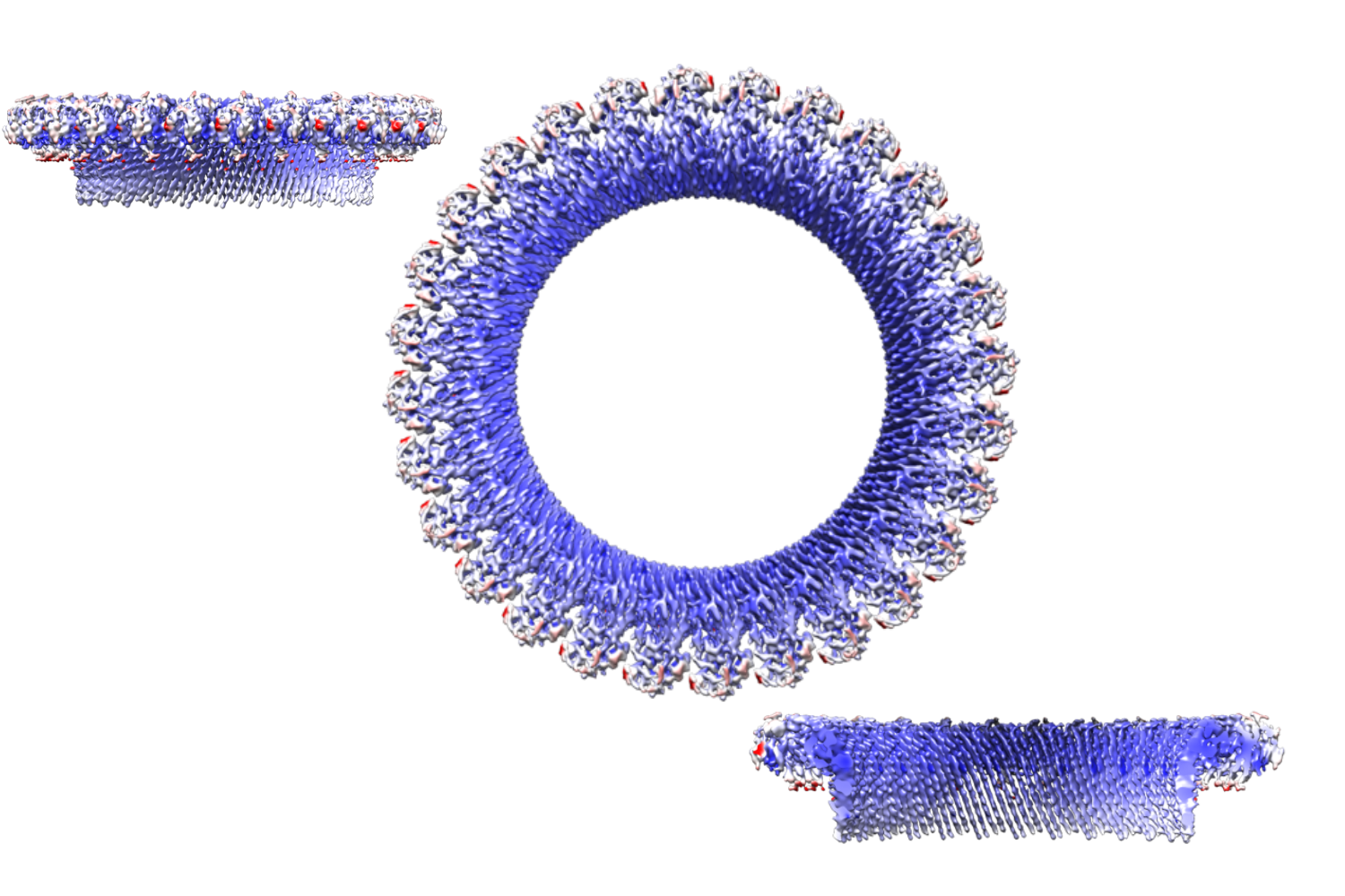
In Ruan’s lab, he is using X-ray crystallography and cryo-electron microscopy (cryo-EM) to understand this process. These techniques allow him to visualize proteins at near-atomic resolution.
Crystallography is useful for studying protein crystals with high stability and homogenous samples. Cryo-EM, on the other hand, constructs images of a protein’s 3D architecture from frozen protein particles, making it more useful for more dynamic, heterogenous samples like those found in immune signaling pathways.
“Cryo-EM allows us to directly visualize how these proteins change shape, assemble, and interact with other molecules, providing mechanistic insight that cannot be obtained from X-ray crystallography, genetics or biochemistry alone,” Ruan says.