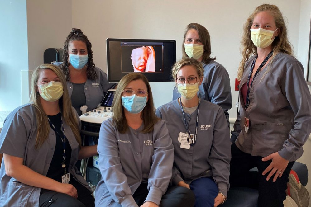
UConn Health’s nearly three dozen ultrasound technologists include diagnostic medical sonographers, vascular sonographers, cardiac sonographers, and perinatal sonographers. All are specially trained on equipment used for imaging and testing to help assess and identify medical conditions.
As Sharie Whittley, one of the diagnostic imaging managers in UConn Radiology describes it, “Diagnostic medical sonography as a specialty is the perfect example of a health care resource shared across all levels of health care delivery and patient care at all ages.”
For Medical Ultrasound Awareness Month, some UConn Health sonographers share some details about their profession and their roles.
Describe your role and how it fits in to the bigger picture of patient care.
Lonnie Collins, vascular technologist, Division of Vascular and Endovascular Surgery: I provide vascular Doppler and ultrasound exams of arteries and veins throughout the body.
Lisa LeBlanc, vascular technologist: I provide vascular sonography images utilizing a combination of gray scale imaging, spectral Doppler waveforms, and color Doppler, of the vasculature within the body for interpretation by our vascular providers here at UConn Health.
Kyla Skoczylas, lead diagnostic medical sonographer, UConn Radiology: Ultrasound is a highly technologist-dependent diagnostic exam that relies on the technologist’s ability to be the eyes of the radiologist. If we do not image something during our scan, the radiologist will not see it either, therefore we must be very skilled in our profession. Sonography is used to help diagnose a patient’s pain and/or other disorders.
Urszula Chmielowiec, lead sonographer, echocardiology lab, Calhoun Cardiology Center: Echocardiogram is a wonderful tool to help in diagnosis of different heart conditions. Can you image looking inside a patient’s heart noninvasively, seeing a valve moving, blood flowing through the chambers, and the human heart beating?
Olena Kiveliyk, lead diagnostic medical sonographer, UConn Health Maternal-Fetal Medicine: A perinatal sonographer specializes in imaging the fetus while in the womb. We look for normal growth and development as well as any abnormalities or pregnancy complications. The role of the perinatal sonographer is to help ensure the fetus and mother are healthy.
Gina Yacovone-Barrett, perinatal sonographer: We work side-by-side with the perinatologists. We specialize in 1st trimester screening exams (to screen for various genetic conditions), detailed fetal anatomy exams (we look from head to toe) and fetal echocardiograms (focus mainly on the fetal heart).
What is unique about your training or your subspecialty within the ultrasound discipline?
Kiveliyk: Our MFM sonographers, Melissa Keeler, Gina Yacovone-Barrett, Samantha Bogner, Deborah Walsh, Ashley Spencer and I, are registered diagnostic medical sonographers in OB-GYN and in addition have special training for the first trimester screening. Melissa, Deborah and I also are trained in fetal echocardiography. All are very talented and kind-hearted sonographers. You have to have a big heart and compassion to provide care to patients in their very special life event.
Skoczylas: During training we were taught all specialties in ultrasound, not just one specialty. In the radiology ultrasound department, we perform a large variety of ultrasound exams. We must be extremely skilled in anatomy and in all areas of sonography.
LeBlanc: I have been fortunate to have had opportunity to be trained on the job in the many sub-specialties (abdominal, obstetrics and gynecology, breast, pediatrics, and vascular) of this great imaging modality right from the start, at the same hospital where I started as a radiologic technologist. Today, most diagnostic medical sonographers need to enter a community college or university-based program to earn an associate’s or bachelor’s degree, or a certificate program after having earned some type of degree. National credentialing is typically required to be employed as a diagnostic medical sonographer, cardiac sonographer, and vascular sonographer/technologist. This credentialing is offered through the ARDMS (American Registry for Diagnostic Medical Sonographers) by passing registry examinations in sonographic physics and instrumentation, and one of the sub-specialties.
What is something most people don’t know about your field?
Collins: I don’t think your average person realizes how many specialties are within the medical ultrasound field, such as general ultrasound, OB/GYN, adult cardiac, pediatric cardiac, muscle/skeletal and vascular.
Skoczylas: Sonography engulfs a large variety of examinations that not many people may be aware of, including OB/GYN, abdominal, small parts, pediatric, breast and vascular. We don’t only scan during pregnancy, which sonography is most known for.
LeBlanc: Maybe some or most people often relate to ultrasound as a job or field that deals with pregnancy and imaging babies in the womb, when this imaging modality offers imaging of much more. Also, ultrasound imaging is extremely safe and patients are not exposed to ionizing radiation. On occasion I will get patients who may think they are having an X-ray, and have to explain that the imaging they are getting is by using sound waves, and not the use of radiation.
What drew you to the profession?
LeBlanc: One of my first jobs in high school was working part-time after school as a radiology transportation aide in a local hospital in my town. This high school job played a significant role in my chosen profession. My work then introduced me to all imaging modalities within radiology. I enjoy working in the health care environment. I truly love helping people. Knowing that my profession and the images that I would acquire during an examination could help diagnose, treat, and possibly save a life was a significant attraction to me.
Deborah Walsh, perinatal sonographer: I was drawn to ultrasound by the amazing ability to noninvasively assess the human body. The moment I had my first experience in a maternal-fetal medicine practice, I knew that was it for me. To be part of such an intimate moment with expectant families is incredibly special and rewarding.
Collins: I was a combat medic and X-ray tech in the Army and I fell in love with helping people.
Chmielowiec: For me it was a very personal decision. My father had a heart condition. I was trying to learn as much as I possibly could about the cardiology field, so that one day I may be able to help him.
Skoczylas: I always wanted to help people and I have always felt the medical field was my calling. I love that sonography gives me the ability to be in direct patient care while providing the radiologist with diagnostic images that can help towards a proper diagnosis for our patients.
How has the pandemic affected your job?
Skoczylas: We have been in the frontlines and have been in full PPE in our patients’ rooms doing many portable ultrasound exams, which can be ergonomically difficult on our bodies, but we do what is best for our patients and anyone else involved. We are all very dedicated to UConn Health and the health of our patients always.
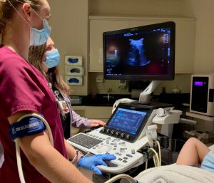
Collins: It definitely has added extra stress that we all are feeling. One thing I have noticed is a heightened need of many patients to connect. Many of my elderly patients have very little socialization now due to the pandemic restrictions and fears.
Melissa Keeler, perinatal sonographer: The COVID boom is a thing; we have been very busy with our high-risk pregnancies. It’s a challenging job but I love every minute.
Kiveliyk: In the fight against COVID-19, perinatal ultrasound is also becoming a front-line tool, since our patients have very limited time frame for their care — no one can prolong pregnancy or postpone delivery. Physicians depend on the highly specialized skills of sonographers to obtain the medical images needed to accurately diagnose and develop the best plan of care in pregnancy. In some COVID-positive patients, whose pregnancies’ had other complications, ultrasound is essential tool to assess for their baby’s well-being during active infection. The number of ultrasounds has not changed during the pandemic. Our pregnant patients had been asked to come to their appointment without a partner. I can’t imagine how nerve-wracking it would have been to attend those ultrasounds solo. Very often we were the first people to share their emotional moments while doing what we do the best: obstetric ultrasound.
Chmielowiec: Driving to work one day with no cars on the road, with no people walking on the streets, made me realize how important the health care profession is, and I was proud to be part of it.
LeBlanc: Limited patient scheduling in the outpatient pavilion during the early onset of the pandemic, and I might say, as with all of us in health care, concern for exposure due to the high risk of contracting COVID-19 before vaccination caused an increase in anxiety and stress for fear of bringing COVID-19 home to our loved ones — lessened, but still present.
What else should we know?
LeBlanc: In addition to its many sub-specialties within, this profession also offers many other career pathways such as teaching, medical research, equipment sales and applications, and employment as a traveling sonographer. The learning in this profession is endless, always something new in equipment applications and imaging techniques in identifying pathology within the body and its vasculature.
Samantha Bogner, perinatal sonographer: It is a special feeling and honor to follow patients through their journey of pregnancy and seeing their baby or babies grow and develop and share that experience with them.
Ashley Spencer, perinatal sonographer: I am excited to be a new team member of the maternal-fetal medicine group it is a great learning environment and an amazing group of Sonographers. There are a lot of opportunities to advance in sonography by providing care to amazing patients.
Yacovone-Barrett: Although highly stressful, it is a very rewarding profession.
Skoczylas: I am proud to be part of the radiology family. We have an amazing team and are all committed to providing our patients with the very best care possible. Many of our sonographers have worked here for a long time! We are happy to be part of the UConn Health family for the long haul.
Collins: Many of our vascular patients return several times and I often develop a friendship. Many will ask me how my dog, Clara, is. Pets and kids and similar life events often weave us together. I do love this profession and I’m one of those people who loves coming to work on Mondays.
Chmielowiec: I would like to take the opportunity to acknowledge and tell all the sonographers in the echo lab how happy I am to be part of this wonderful team. Kim Sheriff, Kristin Marshal, Henry Chua, Lekha Srikanthan, Jessica Dowd, Keti Ademi, Mollie Boucher, and Ewa Kowalczyk. Thank you!
