Researchers have so many ways of probing the unseen. Sound waves whisper of hidden voids, magnetic fields trace fluids flowing through the body. The shape of a crystal reveals the symmetry of its atomic structure, and early cellular biologists derived the existence of organelles from their observations of cell behavior. But there’s nothing quite like seeing, and that’s what makes a microscope so magical.
UConn’s Keramos club asked students to show us what they see when they look through their microscopes. Here’s a sampling of the micrographs submitted, who took them, and what they show.
These first three micrographs were all taken with a Scanning Electron Microscope (SEM). SEMs shoot focused beams of electrons, which either interact with the sample and produce secondary electrons or x-rays, or bounce off the sample and backscatter. The most common type of SEM uses information from the secondary electrons to create the image.

Manganese oxide gets a lot of attention from the material science crowd these days. It’s good at grabbing other molecules onto its surface and oxidizing (adding oxygen to) them in an environmentally benign way, and researchers are working on using it as a catalyst and in fuel cells. Manganese oxide doped with copper is especially good at encouraging molecules with carbon-carbon triple bonds to rearrange their electrons, switching into double-single-double bonds instead. This is handy for making all kinds of useful chemicals. When Biswas and Kriz used a SEM to look at some mesoporous copper-doped manganese oxide they’d synthesized using a new technique, the material was clearly in the shape of nano-sized brains. When they heard of the Keramos competition, they decided to have a little fun, and digitally imposed a nanobrain into a picture of a bell jar.

This image shows two droplets of a platinum salt solution that dried into flower blossom-like structures on the plate. The one on the left kept its shape in the SEM, while the one on the right collapsed. The blossom structure was undesirable for what Jain was trying to look at. But sometimes accidents are beautiful. As Jain says, “This was a bad experiment, but a pretty picture!”

This is a picture of the first sample Deljoo ever collected. It’s octahedral molecular sieve manganese oxide. The sheets of manganese oxide molecules link to make tunnels, a very porous structure with a lot of surface area. She took this image just to get practice with the SEM. When you zoom in, the ‘fur’ on the ‘sea urchins’ is actually plates or rods of manganese oxide. Each sea urchin contains lots and lots of these plates and nanoparticles, which are huge compared to the actual manganese oxide molecules. “Why is it blue? I like blue, and it goes with the sea urchins,” Deljoo says.
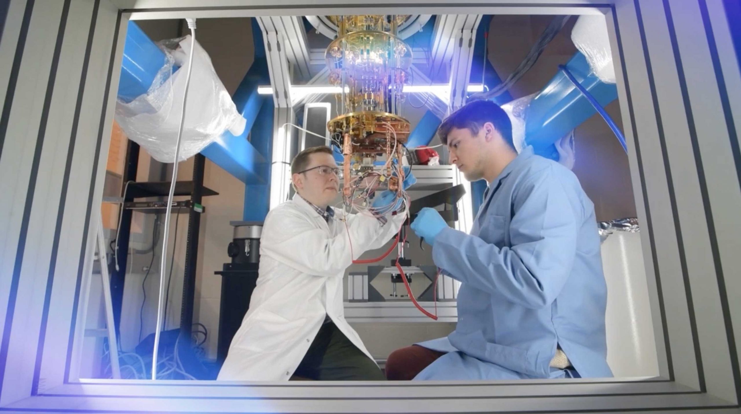
A Transmission Electron Microscope (TEM) uses electrons like an SEM, but it shoots them through an ultra-thin sample. The electrons interact with the sample, altering their amplitude and phase. These changes are then translated into the image we see. In order to work properly, the electrons must be extracted from the very tip of the TEM’s filament. But as the filaments age, they start to flake and splinter and electrons start to shoot off erratically, making them useless.

When Louis Gambino walked into the TEM lab one day to image some samples, he found the TEM’s lanthanum hexaboride filament in the beginning stages of death. So instead of imaging his sample, he took a picture of the filament itself. He calls it My Moon, My Sun. Why? “Because the TEM is my moon and my sun,” Gambino said. A more honest statement of the TEM’s contribution to material science research is seldom heard.

Never let it be said that materials scientists get to have all the fun when it comes to microscopy. MCB graduate student Nicholas Legendre took this picture of a muscle fiber from an injured mouse. Everything that’s green in the picture has expressed a gene, MyoD, involved in muscle regeneration. Legendre wanted to see if adult muscle stem cells could express MyoD and remain distinct, or whether they would inevitably fuse into the muscle. This picture answers that question with a yes! The pinkish-purplish-yellow cell in this image is an adult muscle stem cell that has expressed MyoD after an injury, but still sits separately on the fiber.
Legendre used a confocal microscope to get this shot, which would have been impossible with a regular fluorescent microscope. The confocal microscope uses a pinhole to eliminate out-of-focus light. And unlike a regular optical microscope, it can focus at any depth within a sample, instead of just at the surface.
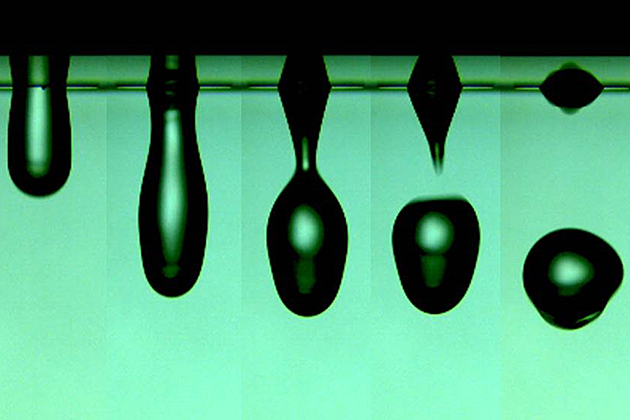
Guo’s micrograph is unique among this group for two reasons. First, it’s not a single picture or series of pictures of the same thing; it’s actually five different shots, each of a different drop of water. Second, the shots were taken with an imaging device made by Guo herself along with a colleague, Brice Bognet. Guo needs to take pictures of complex fluids shooting out of jets, as in an inkjet printer. There are commercially available high-speed cameras that can capture droplets moving that fast, but they’re very expensive and can be blurry. And the inkjetter–the part of the printer that actually shoots out the droplets–can be something of a black box. Guo needs to know exactly how the system works for her research. So she made her own setup on the cheap. She hopes to publish a paper on it later this year.